The following is simply a collection of very interesting research and articles on Graphene and Graphene Oxide. These are excerpts taken from carious articles on the uses and toxicity of Graphene and their effects and uses on and in animals cells. Interestingly at this time – most autopsy analysts will not be able to detect Graphene inside body tissues with their current training and equipment. Add to that the fact they are not primed to look for it.
Terms used; Fetal Bovine Serum = (FBS)
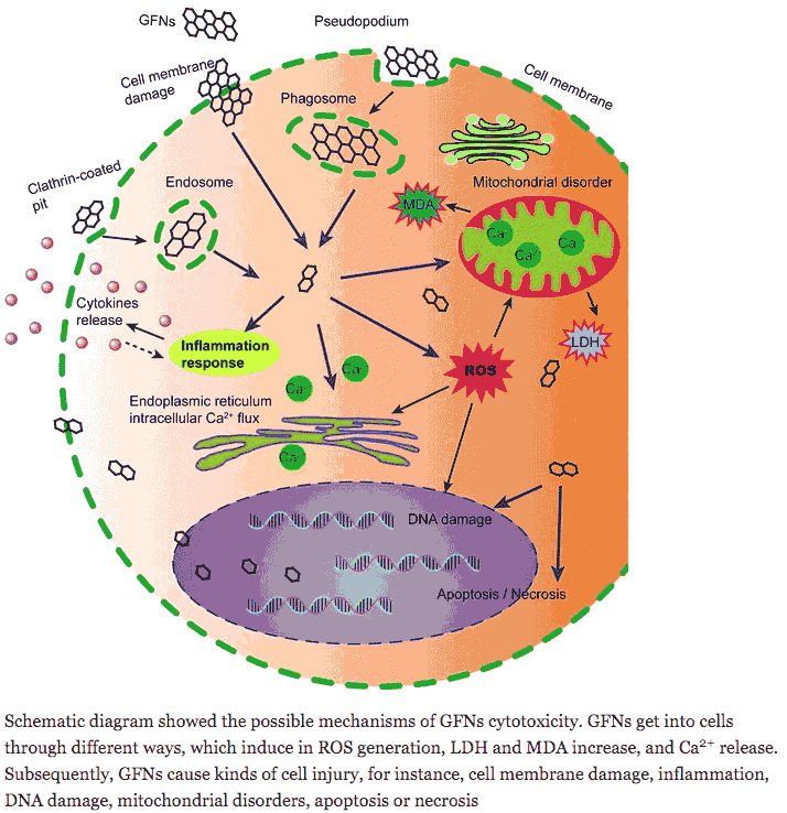
Physical destruction
Graphene is a unique nanomaterial compared with other spherical or one-dimensional nanoparticles due to its two-dimensional structure with sp2-carbons. The physical interaction of graphene nanoparticles with cell membranes is one of the major causes of graphene cytotoxicity [7, 170, 171]. Graphene has high capability to bind with the α-helical structures of peptides because of its favourable surface curvature [172]. At concentration above 75 μg/mL, pristine graphene largely adhered to the surfaces of RAW 264.7 cells and resulted in abnormal stretching of the cell membrane [104]. The strong hydrophobic interactions of GFNs with the cell membrane lead to the morphological extension of F-actin filopodial and cytoskeletal dysfunction. Furthermore, the sharpened edges of GNS may act as ‘blades’, inserting and cutting through bacterial cell membranes [173]. Moreover, GO also damaged the outer membrane of E. coli bacteria directly, resulting in the release of intracellular components [173]. However, TEM imaging revealed that pre-coating GO with FBS eliminated the destruction of cell membranes.
Cytotoxicity of protein corona-graphene oxide nanoribbons on human epithelial cells >>
Abstract
Graphene oxide nanoribbons (GONRs) were synthesized using an oxidative unzipping of multi-walled carbon nanotubes. The interactions of the GONRs with various concentrations of fetal bovine serum or human plasma serum indicated that the GONRs were functionalized substantially by the albumin originated from the two different protein sources. Then, concentration-dependent cytotoxicity of the protein-functionalized GONRs on human epithelial cells was studied. Although the GONRs with concentrations ≤50 μg/mL did not exhibit significant cytotoxicity on the cells (with the cell viability >85%), the concentration of 100 μg/mL exhibited significant cytotoxicity including prevention of cell proliferation and induction of cell apoptosis. These results can provide more in-depth understanding about cytotoxic effects of graphene nanostructures which can be functionalized by the proteins of media.
Graphene-based Materials for Fighting Coronavirus Disease 2019: Challenges and Opportunities
Abstract
The coronavirus disease 2019 (COVID-19) caused by severe acute respiratory syndrome coronavirus 2 (SARS-CoV-2) is considered as serious global threat of this time and greatest challenge for recent days. Several approaches have been carried out in this direction to fight against COVID-19. Among these, nanotechnology is one of the promising approach to face these challenges in the current situation. Recently, graphene-based nanomaterials have been explored for COVID-19 due to its unique physicochemical properties. This mini review provides a recent progress in graphene-based nanomaterials and its applications for diagnosis, detection, decontamination, and protection against COVID-19. Further, main challenges and perspective for fundamental design and development of technologies based on graphene-based materials are discussed and suitable directions to improve these technologies are suggested. This article will provide timely knowledge and future direction about this wonder materials in various biological applications. Read more >>

I love God, Christ, The Bible. art, painting, playing music, improvising music, guitar (first instrument), drumming (not very good at it:), and anything musical or art related. Quick note – my articles are usually spun off in minutes so forgive any errors – hopefully you get the essence of the article and I’ll pick up mistakes later. If you want to write of 101ChristianMagazine and inspire and edify Christians with articles on faith, general news, hobbies or anything that interests you email me at: epgb101@gmail.com:)


